Showing posts with label invention. Show all posts

Tuesday, August 8, 2023

The invention of Lasik eye surgery by Gholam Peyman

In 1948, Father Szuniewicz, recognized as a trailblazer in the field of refractive surgery, voyaged to the United States with the intention of advancing his investigation into corneal reshaping. He embarked on this pursuit at Yale University in New Haven, CT.

Starting as early as the 1950s, medical researchers began delving into techniques to correct vision by manipulating the cornea's shape. However, it was ophthalmologist Jose Barraquer from Colombia who initiated the historical trajectory of LASIK by devising the initial surgical remedy for vision correction. He honed a method that involved fashioning a flap within the cornea to facilitate alterations in its shape.

Dr. Gholam Peyman initially undertook the study and evaluation of the potential utility of a CO2 laser to modify the structure of rabbit eye corneas. Nonetheless, he found this laser to be inadequate for the required procedure and lacking in its ability to prevent subsequent scarring.

Peyman's primary aim was to avoid surface ablation in order to sidestep potential corneal scarring and the accompanying discomfort resulting from the removal of the corneal epithelium, a step needed to expose the cornea's surface.

In 1980, Peyman published an article outlining the feasibility of performing eye surgery using ablative laser technology. However, this concept remained speculative at that time due to the unavailability of the necessary laser advancements.

During the 1980s, Rangaswamy Srinivasan, an IBM researcher stationed in Yorktown Heights, NY, delved into the interaction of laser technology with organic matter, particularly exploring the impacts of ultraviolet light. A significant breakthrough occurred in 1980 when Dr. Srinivasan confirmed that an excimer laser could extract living tissue without causing thermal harm to surrounding cells.

Towards the end of 1982, he stumbled upon an article from IBM Laboratories published in Laser Focus, which detailed their work involving excimer lasers and organic materials.

Consequently, in July 1985, he submitted a patent application describing a technique for altering corneal refractive errors through laser ablation beneath a corneal flap. This patent was eventually granted in June 1989 after two revisions. Peyman initiated a series of experimental studies in collaboration with the Physics Department of the University of Helsinki, Finland, to assess the effects of various excimer lasers.

In 1990, European eye specialists Ioannis Pallikaris and Lucio Burrato pioneered a method that laid the foundation for LASIK surgery. Termed the "flap and zap" technique, it encompassed the creation of a corneal flap, reshaping tissue using a laser, and subsequently sealing the flap to promote healing.

Maintaining awareness of the potential limitations of his innovation, Peyman dedicated substantial time and effort in the following years to enhancing them. In a bid to optimize the risk-to-benefit ratio of the LASIK procedure, he introduced and obtained patents for a comprehensive range of ablative and non-ablative inlays in 2004. These were designed to be placed beneath the surgically fashioned corneal flap.
The invention of Lasik eye surgery by Gholam Peyman

Thursday, November 3, 2022

The invention of cataract laserphaco probe

In 1988, Patricia Era Bath patented the cataract laserphaco probe that helped save the eyesight of a million. Patricia Bath (November 4, 1942 – May 30, 2019) was the first African American to complete a residency in ophthalmology and the first African American female doctor to receive a medical patent.

Bath was born in Harlem, New York, to Rupert and Gladys Bath. After graduating from high school in only two years, Bath headed to Hunter College, where she earned a bachelor's degree in 1964. She received her medical degree from Howard University College of Medicine in Washington, D.C., interned at Harlem Hospital from 1968 to 1969, and completed a fellowship in ophthalmology at Columbia University from 1969 to 1970.

Following her fellowship in ophthalmology at Columbia University, from 1970 to 1973, she completed her training at New York University, where she became the first Black resident in ophthalmology. In 1974 Bath joined the faculty of UCLA and Charles R. Drew University as an assistant professor of surgery (Drew) and ophthalmology (UCLA).

In 1981, Bath began working on her most well-known invention: the Laserphaco Probe (1986). Harnessing laser technology, the device created a less painful and more precise treatment of cataracts. The laserphaco probe is a tool that uses a tiny laser to safely vaporize cataracts in a patient’s eye. Once the probe has removed the cataract, a surgeon is then able to remove the lens of the eye and insert a replacement.

It took her nearly five years to complete the research and testing needed to make it work and apply for a patent. She received a patent for the device in 1988, becoming the first African American female doctor to receive a patent for a medical purpose.
The invention of cataract laserphaco probe

Monday, May 16, 2022

History of computed tomographic (CT) scanner

In the early 1900s an Italian radiologist named Alessandro Vallebona invented tomography which used radiographic film to see a single slice of the body. As conventional tomography evolved, it was still considered ineffective when it came to imaging soft tissues.

The computed tomographic (CT) scanner was conceived in 1967 by British engineer Sir Godfrey Hounsfield at EMI Central Research Laboratories using x-ray technology. The first patient studies were performed on Friday, October 1, 1971 in Wimbledon, England but it was not publicized until a year later.

Sir Godfrey Hounsfield continued to improve the quality of the devise and the first commercially available CT scanner was available in 1972. He co-invented the technology with physicist Dr. Allan Cormack.

However, it was the mathematical theory of Johann Radon way back in 1917, called “Radon transform,” that brought the technology to life. He demonstrated that the image of a three-dimensional object can be constructed from an infinite number of two-dimensional images of the object.

Another mathematical advancement that Hounsfield built on is the “Algebraic Reconstruction Technique,” which was formulated by Polish mathematician Stefan Kaczmarz in 1937. Stefan Kaczmarz proposed a simple method, called the Kaczmarz algorithm, to solve iteratively systems of linear equations Ax = b in Euclidean spaces. This procedure employs cyclic orthogonal projections onto the hyperplanes associated with such a system.

Both theories by Johann Radon and Stefan Kaczmarz were adopted by Hounsfield to create one of the greatest advancements in medical history.

In 1973, the first CT scanners were installed in the United States. Sir Hounsfield would go on to share the 1979 Nobel Prize in Physiology or Medicine with Allan M Cormack.
History of computed tomographic (CT) scanner

Saturday, February 5, 2022

History of ultrasound in medicine

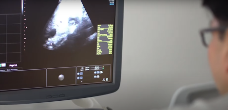
Lazzaro Spallanzani an Italian physicist is most often credited person for discovering ultrasonography. In 1794, he analyzed the basic mechanisms of the navigation of flying bats in the dark, and rightly deduced that bats employed sound rather than light to orient themselves.

In 1877, Jacques and Pierre Currie were the first to discover piezoelectricity. This discovery was very important to ultrasound since the ultrasound transducers or probes receive and emit sound waves by using the piezoelectric effect.

Powerful high frequency ultrasonic echo-sounding device was developed by emminent French physicist Paul Langévin and Russian scientist Constantin Chilowsky, then residing in France.

In 1915, Paul Langévin, commissioned to create a device that would detect objects found on the bottom of the ocean. He ended up inventing a hydrophone, which was referred to as the “first transducer” by the World Congress on Ultrasound in Medical Education.

In the 1920s and 1930s, ultrasound was used for physical therapy, primarily for members of Europe’s soccer teams. It also was used for sterilization of vaccines and for cancer therapy in combination with radiation therapy.

Karl Dussik, a neurologist and a psychiatrist at the University of Vienna in Austria, is generally regarded as the first physician to employ ultrasound in medical diagnosis in 1947-1948. He and his brother Friederick, a physicist, introduced hyperphonography, a technique which used ultrasound to visualize the cerebral ventricles. They transmitted an ultrasound beam through the human skull in attempts of detecting brain tumors.

In 1949 Douglas Howry, a radiologist and coworkers used surplus radio and Air Force and radar parts to build a pulse-echo ultrasonic scanner capable of making two-dimensional images. In 1950, using a 35-mm camera, Howry recorded the first cross-sectional images with ultrasound.

In United States, George Ludwig, while serving at the Naval Medical Research Institute in Maryland first applied ultrasound for medical purposes in the late 1940s. In 1949, he carried out research into gallstones embedded in soft tissues, using a through transmission technique. His pioneering investigations into the interactions between ultrasonic waves and animal tissues, helped lay the foundations for the later successful use of ultrasound in medical practice.

In United Kingdom, in 1956, Dr. Ian Donald, an English physician, incorporated ultrasound into the OB/GYN field of medicine. Ian Donald and an engineer named Tom Brown developed the first prototype system for ultrasound, but it wasn’t perfected until the end of the 1950s.

In the 1980s, ultrasound technology became more sophisticated with improved image quality and 3D imaging capabilities. These improvements continued into the 1990s with the adoption of 4D (real time) capabilities.
History of ultrasound in medicine

Tuesday, January 18, 2022

Invention of stethoscope

Ebers Papyrus (1500 BCE -Before Common Era) and the Vedas of Ancient India around 1400 BCE are thought to have the earliest recorded reference to breath sounds (sound inside human breathing organ - the lungs).

Joseph Leopold Auenbrugger (1722-1809) is considered to be the father of the modern physical examination, which is based on percussion. Percussion of the chest was used regularly during the 18th century to reveal various sounds and to help outline enlarged organs or lesions in the chest.

Percussion Leopold Auenbrugger performed by knocking directly on the thorax with the finger or cupped hands. His discovery of the percussion sounds from the chest during inhaling and exhaling originates from his work in 1760 at the Vienna Military Hospital.

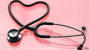
The word "stethoscope" originates from the Greek words "stetho" meaning "chest" and "scopein" meaning to view or observe.

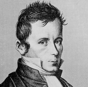
The stethoscope was invented by the French physician Rene Theophile Hyacinthe Laënnec (1781-1826), a student of Corvisart and later physician to the famous Saltpetre and Necker Hospital in Paris. It was a unitubular cyclinder first made of paper and pasteboard in 1819 and later turned into wooden tube. He named it "LE CYCLINDER". He believed a method to diagnose chest conditions was needed, particularly for stout individuals where direct auscultation to the chest was either inadequate or embarrassing, especially for his female patients.
Laënnec was a military surgeon during the Revolution and Napoleon era. He studied medicine in Paris with the great physicians Bichat and Corvisart. After graduation, he worked in the prestigious Paris hospitals Beaujon and Necker. He got a reputation of skilled doctor and had many clients.

In 1819 he summarized his studies with the stethoscope in a two-volume work on more than 900 pages. In those days, wooden stethoscopes made according to his instructions were sold in Piccadilly, London, for four shillings each. It was monaural and could be applied to one ear only.

Laennec had a special interest in diseases of the chest because tuberculosis was rampant during the 18th century; his own mother died of the disease. Laënnec's invention of the stethoscope originated from the difficulty of examining a fat young woman laboring under general symptoms

of heart problems. He hesitated to put his ear directly onto her chest, because she was a young girl. Therefore, during the examination, he rolled a piece of paper into a tube and placed one end of the tube on the girl's chest and the other end against his ear. The inspiration for this is said to be some children's play with wooden pieces that reinforced the sound of a scraper at the other end of the tree. To his delight, Laënnec discovered at finding that he could perceive sounds clearly and distinctly.

He continued his experiments and soon found that an aperture instead of solid wood was even better and that pine was the wood most suitable. This caused him to develop a tube in wood - the first monaural stethoscope.

His first stethoscope design was approximately 30 cm long, 2 cm in diameter, and cylindrical. He made it in three parts with a funnel-shaped section at the bell end. Laënnec stated that the complete device is best to hear heart sounds while the respiratory sounds are heard most clearly when the small funnel is removed.

James Hope (1801-1841) a medical graduate of Edinburgh University, and later physician to St. George’s Hospital, London, who spent some time in Laënnec’s clinic in Paris is credited with having influenced the acceptance of the stethoscope by the London medical community.

In 1826, the stethoscope was employed almost universally in France and extensively in Germany and less frequently in England. It was probably first used in the United States of America around 1821-1823.
Invention of stethoscope

Tuesday, July 19, 2016

History of Vicks VapoRub

Lunsford Richardson sold his drugstore and moved to Greensboro with wife, Mary Lynn and their three children in 1891.

He then purchased the Porter Drug Store at 121 South Elm Street.  It served as a popular spot to socialize as well as shop. In 1905 at age fifty, he left the wholesale business and with $8,000 capital launched the wholly owned Vick Family Remedies Companies.

Lunsford mostly found his passion in developing home remedy, creating his own formulas to help customers relive headaches, coughs, and other common ailments.

He experimented with a new remedy to ease his children’s croup symptoms in 1894, blending an ointment of menthol, eucalyptus and other natural ingredients.

Richardson blended menthol into petroleum jelly and produces Richardson’s Coup and Pneumonia Cure Salve.

When rubbed on the chest the stuff did produce the sensation of heat, and as the menthol evaporated it did clear the nasal passages.

The products name was too long, however, so Richardson christened his line Vick Family Remedies, borrowing the name from his brother–in-law, Dr. Joshua Vick. The name Vicks was short, catchy and easy to remember.

In 1917 the company began a widespread distribution of samples on a house to house basis in many areas of the Northeast. Vick was probably the first drug company to hire a doctor to review the medical accuracy of all its advertising copy.

Sales increased each year and by 1929, Vicks products could be purchased in sixty foreign countries.

In 1938 the Richardson acquired the William S. Merrel Company. From this small town operation, Richardson Merrell grew into multi-national $500 million enterprise.

In 1956 Bendectin was introduced to the market, it was a product of Richardson Merrell, whose parent was Vicks.

In October 1985, the board approved the friendly sale to Procter & Gamble, securing a more favorable stock price and outcome for employees.
History of Vicks VapoRub

Saturday, December 13, 2014

First artificial limbs for soldiers

Surgeons performed more than 50,000 amputations during the Civil War and nearly 75 percent of the patients survived.

In 1862, the government provided the amputee veteran with $75 for an artificial leg and $50 for an artificial arm. By 1895 the US government had granted 144 patents for legs and in 1917, as the United States entered World War I, there were 200 artificial limb manufacturers in the country.

Among the notable manufacturers of artificial limbs was B. Franklin Palmer of Philadelphia, himself an amputee, who had been making such limbs since well before the Civil War.

Ambroise Pare (1509-1590) was the first to tackle the problem of producing artificial limbs and the first to establish their feasibility.

He joined the French army and became a military surgeon in 1536, and consequently was surrounded by the need for these aids. He became a leading surgeon of the Renaissance at a time when surgeons were still considered on a par with butchers.

His inventions were mainly designed for wounded soldiers and he devised for them several arms and hands that were successful.

In his books Oeuvres, he described and provided drawings in great details of an artificial hand, arm and leg designed for soldiers.
First artificial limbs for soldiers

The most popular articles

Selected Articles